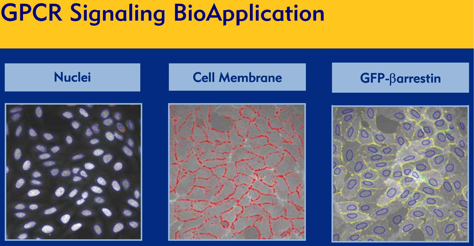
Demonstration of the GPCR Signaling
BioApplication utilizing Transfluor® technology.
Cells expressing both GFP-βarrestin (right) and the
β2-adrenergic receptor were stained with Hoechst
nuclear dye (left) and a red fluorescent cell
membrane marker (center). Images of all three
fluorescent channels were automatically acquired
on an ArrayScan® HCS Reader and analyzed
using the GPCR Signaling BioApplication.

Nuclei
(blue overlays) and membranes (red spots) are
identified automatically, and the distribution of the
GFP-βarrestin fusion (yellow and green spots) is
determined. In this example of early activation
the receptor is mostly in small pits co-localized
with the membrane.